This site uses cookies to improve your experience. To help us insure we adhere to various privacy regulations, please select your country/region of residence. If you do not select a country, we will assume you are from the United States. Select your Cookie Settings or view our Privacy Policy and Terms of Use.
Cookie Settings
Cookies and similar technologies are used on this website for proper function of the website, for tracking performance analytics and for marketing purposes. We and some of our third-party providers may use cookie data for various purposes. Please review the cookie settings below and choose your preference.
Used for the proper function of the website
Used for monitoring website traffic and interactions
Cookie Settings
Cookies and similar technologies are used on this website for proper function of the website, for tracking performance analytics and for marketing purposes. We and some of our third-party providers may use cookie data for various purposes. Please review the cookie settings below and choose your preference.
Strictly Necessary: Used for the proper function of the website
Performance/Analytics: Used for monitoring website traffic and interactions
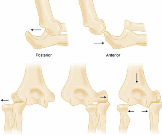
PMID: 32644703 Robinson PM, Griffiths E, Watts AC. PMID: 27227986 Glover NM, Black AC, Murphy PB. PMID: 31082090 Post Peer Reviewed By: Anand Swaminathan MD, MPH (Insta @EMSwami) The post Elbow Dislocations appeared first on REBEL EM - Emergency Medicine Blog. Treasure Island (FL): StatPearls Publishing; 2024 Jan–. 2023 Nov 5.
This dynamic change is diagnostic of ACS. Cardiology was consulted and agreed that his history was high risk for ACS and a next-day angiogram was merited. This was also non-diagnostic for OMI, although the dynamic changes are diagnostic of ACS. ECG at time 82 minutes: What do you think?
Paper: Alwang AK, Law AC, Klings ES, Cohen RT, Bosch NA. PMID: 28106307 Alwang AK, Law AC, Klings ES, Cohen RT, Bosch NA. appeared first on REBEL EM - Emergency Medicine Blog. To assess the clinical impact and relevance of these concerns, Alwang et al. performed the retrospective cohort study below ( Alwang 2024 ).
3) ACS with possible additional ischemia from atrial fib with RVR 4) Hemorrhage/dehydration/sepsis/etc., I was fairly certain that this was a type II (demand ischemia) MI and that this patient was not having ACS. He did not have ACS. Not all myocardial infarction is due to ACS 2. He was treated for urosepsis and did well.
ACS is dynamic. interesting spontaneous reperfusion case 1413140 prehospital STEMI first ED ECG is here, with 3/10 pain: But this is the same patient just 10 minutes before, with 7/10 pain Isn't it ridiculous to say that the patient has both a STEMI and an NSTEMI? It can't be given one static name. Now the patient has one disease: OMI.
World Bank Blogs. link] Hummell AC, Cummings M. A qualitative study on African immigrant and refugee families experiences of accessing primary health care services in Manitoba, Canada: its not easy! Int J Equity Health. 2017;16(1):5. doi:10.1186/s12939-016-0510-x Verme P. March 28, 2023. Accessed October 5, 2024. 2022;37(1):41-49.
Full blog post here. PMID: 39461792 Bottom line: The WOMAN 2 trial is a large double-blind RCT that shows no benefit of TXA in the prevention of postpartum hemorrhage, which fits with all of the existing literature demonstrating no role for TXA in the management of postpartum hemorrhage. Emerg Med J. 2019 Jan;36(1):2-3. Epub 2018 Oct 25.
Translate Follow us on X (Twitter) Follow @smithECGBlog Follow @PendellM Follow @ekgpress Follow @AslangerE Follow @ecgcases Follow @PMcardioBot Total Pageviews Use this Blog as a Textbook I highly recommend using this blog as an atlas or textbook. Five Primary Patterns of Ischemic ST depression, without ST elevation.
It is important to note that these findings, if due to atherothrombotic acute coronary syndrome (ACS), are NOT due to occlusion of the left main, as is frequently stated in online postings and in literature. It is most commonly due to demand ischemia, not due to ACS! If it were ACS, what reperfusion options were available?
One must remember that acute pulmonary edema is frequently triggered by ACS. In this case, it is reasonable to assume that there is no ACS. Here is the troponin profile: Troponins this high are almost always due to type 1 MI (ACS, plaque rupture) This was a surprise. Severe, life threatening ACS may have no ST Segment shifts.
May mimic ACS Lesson : 1. And such absence may or may not lead one to pursue other diagnoses. Remember that Pulmonary Embolus: 1. Causes anterior T-wave inversion plus T-wave inversion in III 2. Often has elevated troponin 3. Often has elevated BNP 4. What appears to be pulmonary edema on chest X-ray may, in fact, be infarct or infiltrate.
The history is highly suggestive of ACS. The patient was given a diltiazem bolus and drip, her pulse slowed, and her chest pain completely resolved. Another ECG was recorded: Atrial Fib with a controlled rate The ST Depression is mostly resolved with this slower rate The first troponin was, not surprisingly, elevated at 1.07
This blog post was written to accompany a Simulated PEM Adventure at Neptune 2025, the UK Paediatric Trauma Conference. Govindraj R, Binda DD, Harris AC et al. Let’s be honest: most of us have had moments at work where we’ve felt hurt by something someone has said or done. Healthcare can be high-pressure and emotionally demanding.
One study found that the best discrimination of stress cardiomyopathy from ACS was possible with the ratio of NT-proBNP/cTnT on the 2nd day. and an accuracy of ∼96% in detecting stress cardiomyopathy as opposed to ACS. NT-proBNP and CTnT in ACS and Takotsubo) Smith : However, this is not of any help with the acute diagnosis!
You can find more details in the full blog post. PECARN looks at probiotics for toddlers diarrhea… Schnadower D, Tarr PI, Casper TC, Gorelick MH, Dean JM, O’Connell KJ, Mahajan P, Levine AC, Bhatt SR, Roskind CG, Powell EC, Rogers AJ, Vance C, Sapien RE, Olsen CS, Metheney M, Dickey VP, Hall-Moore C, Freedman SB.
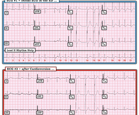
Learning Point: When there is ST depression and atrial fibrillation with RVR, it is very useful to cardiovert if possible, or slow AV conduction, before assuming that the ST depression is a result of ACS. == MY Comment by K EN G RAUER, MD ( 6/16/2020 ): == Interesting case presented by Dr. Smith — regarding this 60-something year old man who presented (..)
When seeing a South Asian patient with chest pain, concern for ACS must be heightened, given their disproportionately higher risk of CAD, despite often lacking traditional risk factors.) Patient initially presented at 9 PM to a referring facility with hsTnI 13 (ref: < 34 ng/L) then 30, then 60.
Translate Follow us on X (Twitter) Follow @smithECGBlog Follow @PendellM Follow @ekgpress Follow @AslangerE Follow @ecgcases Follow @PMcardioBot Total Pageviews Use this Blog as a Textbook I highly recommend using this blog as an atlas or textbook. Five Primary Patterns of Ischemic ST depression, without ST elevation.
Translate Follow us on X (Twitter) Follow @smithECGBlog Follow @PendellM Follow @ekgpress Follow @AslangerE Follow @ecgcases Follow @PMcardioBot Total Pageviews Use this Blog as a Textbook I highly recommend using this blog as an atlas or textbook. Five Primary Patterns of Ischemic ST depression, without ST elevation.
Translate Follow us on X (Twitter) Follow @smithECGBlog Follow @PendellM Follow @ekgpress Follow @AslangerE Follow @ecgcases Follow @PMcardioBot Total Pageviews Use this Blog as a Textbook I highly recommend using this blog as an atlas or textbook. Five Primary Patterns of Ischemic ST depression, without ST elevation.
Translate Follow us on X (Twitter) Follow @smithECGBlog Follow @PendellM Follow @ekgpress Follow @AslangerE Follow @ecgcases Follow @PMcardioBot Total Pageviews Use this Blog as a Textbook I highly recommend using this blog as an atlas or textbook. Five Primary Patterns of Ischemic ST depression, without ST elevation.
They show that if there is not >/= 1 mm STE in aVR, then ACS is highly unlikely to be due to severe 3-Vessel disease or Left Main. An early and simple predictor of severe left main and/or three-vessel disease in patients with non-ST-segment elevation acute coronary syndrome. Am J Cardiol;107(4):495-500. why is this important?
The ED physicians were of course worried about ACS, and they obtained these POCUS echos: Apical 4 chamber Overall LV function is moderately decreased Parasternal short axis: Is there an anterior wall motion abnormality? Parasternal long axis: Again, decreased LV function 101.8 WBC 27,000 0.299 trop UA with infection lactate 3.4
Translate Follow us on X (Twitter) Follow @smithECGBlog Follow @PendellM Follow @ekgpress Follow @AslangerE Follow @ecgcases Follow @PMcardioBot Total Pageviews Use this Blog as a Textbook I highly recommend using this blog as an atlas or textbook. Five Primary Patterns of Ischemic ST depression, without ST elevation.
Translate Follow us on X (Twitter) Follow @smithECGBlog Follow @PendellM Follow @ekgpress Follow @AslangerE Follow @ecgcases Follow @PMcardioBot Total Pageviews Use this Blog as a Textbook I highly recommend using this blog as an atlas or textbook. Five Primary Patterns of Ischemic ST depression, without ST elevation.
Learning point: ST Depression maximal in leads V1-V4 in the context of high suspicion for ACS is posterior OMI until proven otherwise, because subendocardial ischemia manifests with STD maximal in V5-6. Formal bubble contrast echo the next day was completely normal, as expected. This is true unless: 1.
Translate Follow us on X (Twitter) Follow @smithECGBlog Follow @PendellM Follow @ekgpress Follow @AslangerE Follow @ecgcases Follow @PMcardioBot Total Pageviews Use this Blog as a Textbook I highly recommend using this blog as an atlas or textbook. Five Primary Patterns of Ischemic ST depression, without ST elevation.
Translate Follow us on X (Twitter) Follow @smithECGBlog Follow @PendellM Follow @ekgpress Follow @AslangerE Follow @ecgcases Follow @PMcardioBot Total Pageviews Use this Blog as a Textbook I highly recommend using this blog as an atlas or textbook. Five Primary Patterns of Ischemic ST depression, without ST elevation.
Translate Follow us on X (Twitter) Follow @smithECGBlog Follow @PendellM Follow @ekgpress Follow @AslangerE Follow @ecgcases Follow @PMcardioBot Total Pageviews Use this Blog as a Textbook I highly recommend using this blog as an atlas or textbook. Five Primary Patterns of Ischemic ST depression, without ST elevation.
Translate Follow us on X (Twitter) Follow @smithECGBlog Follow @PendellM Follow @ekgpress Follow @AslangerE Follow @ecgcases Follow @PMcardioBot Total Pageviews Use this Blog as a Textbook I highly recommend using this blog as an atlas or textbook. Five Primary Patterns of Ischemic ST depression, without ST elevation.
EDACS is broader—captures symptoms suspicious for ACS, not just chest pain, and performs well in low-risk patients. Clinical Bottom Line Both HEART and EDACS are best utilized for identifying patients with suspected ACS who are at low risk for MACE. Cardiovascular Read More REBEL Cast Ep114: High Flow O2, Suspected ACS, and Mortality?
Translate Follow us on X (Twitter) Follow @smithECGBlog Follow @PendellM Follow @ekgpress Follow @AslangerE Follow @ecgcases Follow @PMcardioBot Total Pageviews Use this Blog as a Textbook I highly recommend using this blog as an atlas or textbook. Five Primary Patterns of Ischemic ST depression, without ST elevation. Acute aphasia.
Translate Follow us on X (Twitter) Follow @smithECGBlog Follow @PendellM Follow @ekgpress Follow @AslangerE Follow @ecgcases Follow @PMcardioBot Total Pageviews Use this Blog as a Textbook I highly recommend using this blog as an atlas or textbook. Five Primary Patterns of Ischemic ST depression, without ST elevation.
Translate Follow us on X (Twitter) Follow @smithECGBlog Follow @PendellM Follow @ekgpress Follow @AslangerE Follow @ecgcases Follow @PMcardioBot Total Pageviews Use this Blog as a Textbook I highly recommend using this blog as an atlas or textbook. Five Primary Patterns of Ischemic ST depression, without ST elevation.
Translate Follow us on X (Twitter) Follow @smithECGBlog Follow @PendellM Follow @ekgpress Follow @AslangerE Follow @ecgcases Follow @PMcardioBot Total Pageviews Use this Blog as a Textbook I highly recommend using this blog as an atlas or textbook. Five Primary Patterns of Ischemic ST depression, without ST elevation.
Translate Follow us on X (Twitter) Follow @smithECGBlog Follow @PendellM Follow @ekgpress Follow @AslangerE Follow @ecgcases Follow @PMcardioBot Total Pageviews Use this Blog as a Textbook I highly recommend using this blog as an atlas or textbook. Five Primary Patterns of Ischemic ST depression, without ST elevation.
Translate Follow us on X (Twitter) Follow @smithECGBlog Follow @PendellM Follow @ekgpress Follow @AslangerE Follow @ecgcases Follow @PMcardioBot Total Pageviews Use this Blog as a Textbook I highly recommend using this blog as an atlas or textbook. Five Primary Patterns of Ischemic ST depression, without ST elevation.
He was started on ACS therapy and loaded with Plavix. This procedures were done due to prior similar presentations and concern of ACS. This is the most likely caused of his fixed ST elevation and no concern for ACS. Initial ECG showed ST elevation with progression of biphasic T wave concerning for reperfusion injury.
Translate Follow us on X (Twitter) Follow @smithECGBlog Follow @PendellM Follow @ekgpress Follow @AslangerE Follow @ecgcases Follow @PMcardioBot Total Pageviews Use this Blog as a Textbook I highly recommend using this blog as an atlas or textbook. Simple ACS? Some are STEMI-equivalents. A male in his 50s with chest pain.
Translate Follow us on X (Twitter) Follow @smithECGBlog Follow @PendellM Follow @ekgpress Follow @AslangerE Follow @ecgcases Follow @PMcardioBot Total Pageviews Use this Blog as a Textbook I highly recommend using this blog as an atlas or textbook. Five Primary Patterns of Ischemic ST depression, without ST elevation.
Translate Follow us on X (Twitter) Follow @smithECGBlog Follow @PendellM Follow @ekgpress Follow @AslangerE Follow @ecgcases Follow @PMcardioBot Total Pageviews Use this Blog as a Textbook I highly recommend using this blog as an atlas or textbook. Five Primary Patterns of Ischemic ST depression, without ST elevation.
Translate Follow us on X (Twitter) Follow @smithECGBlog Follow @PendellM Follow @ekgpress Follow @AslangerE Follow @ecgcases Follow @PMcardioBot Total Pageviews Use this Blog as a Textbook I highly recommend using this blog as an atlas or textbook. Five Primary Patterns of Ischemic ST depression, without ST elevation.
Translate Follow us on X (Twitter) Follow @smithECGBlog Follow @PendellM Follow @ekgpress Follow @AslangerE Follow @ecgcases Follow @PMcardioBot Total Pageviews Use this Blog as a Textbook I highly recommend using this blog as an atlas or textbook. Five Primary Patterns of Ischemic ST depression, without ST elevation.
We organize all of the trending information in your field so you don't have to. Join 5,000+ users and stay up to date on the latest articles your peers are reading.
You know about us, now we want to get to know you!
Let's personalize your content
Let's get even more personalized
We recognize your account from another site in our network, please click 'Send Email' below to continue with verifying your account and setting a password.


Let's personalize your content