This site uses cookies to improve your experience. To help us insure we adhere to various privacy regulations, please select your country/region of residence. If you do not select a country, we will assume you are from the United States. Select your Cookie Settings or view our Privacy Policy and Terms of Use.
Cookie Settings
Cookies and similar technologies are used on this website for proper function of the website, for tracking performance analytics and for marketing purposes. We and some of our third-party providers may use cookie data for various purposes. Please review the cookie settings below and choose your preference.
Used for the proper function of the website
Used for monitoring website traffic and interactions
Cookie Settings
Cookies and similar technologies are used on this website for proper function of the website, for tracking performance analytics and for marketing purposes. We and some of our third-party providers may use cookie data for various purposes. Please review the cookie settings below and choose your preference.
Strictly Necessary: Used for the proper function of the website
Performance/Analytics: Used for monitoring website traffic and interactions
The T wave changes that have occurred are widespread, and not in a typical coronary distribution. One study found that the best discrimination of stress cardiomyopathy from ACS was possible with the ratio of NT-proBNP/cTnT on the 2nd day. and an accuracy of ∼96% in detecting stress cardiomyopathy as opposed to ACS.
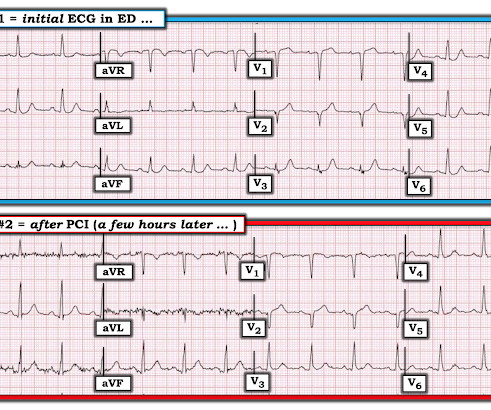
A 41-year-old South Asian male with history of hypertension, alcohol use disorder and hyperlipidemia, who has a strong family history of CAD presented with central substernal burning, pressure, and pain with associated diaphoresis. Coronary angiography before and after intervention is shown below. New PMcardio for Individuals App 3.0
An early and simple predictor of severe left main and/or three-vessel disease in patients with non-ST-segment elevation acute coronary syndrome. They show that if there is not >/= 1 mm STE in aVR, then ACS is highly unlikely to be due to severe 3-Vessel disease or Left Main. Am J Cardiol;107(4):495-500. why is this important?
I think the right answer is that the patient probably needs emergent angiography to rule out acute coronary occlusion, but because it is such a complicated patient with such atypical symptoms, it is best to consult with cardiology about the case before activating. There was clearly a myocardial infarction and severe coronary disease.
There is increased LV cavity dimensions with an increase in transient ischemic dilation, suggesting Left Main, or 3-vessel coronary artery disease. 2. Coronary angiography reveals significant and severe CAD involving all three epicardial vessels. Strongly positive stress ECG Lexiscan administration. Type I ischemia.
He denied any known history of CAD, but did report ASCVD risk factors to include HTN, HLD, and DM. I interpreted the ECG as VT with two primary etiological possibilities: 1. Abrupt plaque ulceration of Type 1 ACS leading to VT. 2. Here is the ECG after 200J.
Major adverse cardiac event rates in moderate-risk patients: Does prior coronary disease matter? Major adverse cardiac event rates in moderate-risk patients: Does prior coronary disease matter? He has no history of coronary artery disease. If we thought about ACS, we brought them in. AEM June 2022. AEM June 2022.
A 63 year old man with a history of hypertension, hyperlipidemia, prediabetes, and a family history of CAD developed chest pain, shortness of breath, and diaphoresis after consuming a large meal at noon. They too have dense white masses consistent with coronary atherosclerosis. Edited by Smith He also sent me this great case.
Moreover, he had no pertinent medical history to report in terms of CAD, HTN, HLD, or DM, for example. Although the attending crews did not consider the ECG pathognomonic for occlusive thrombosis, they nonetheless considered the patient high-risk for ACS and implored him to reconsider. A 12 Lead ECG was recorded.
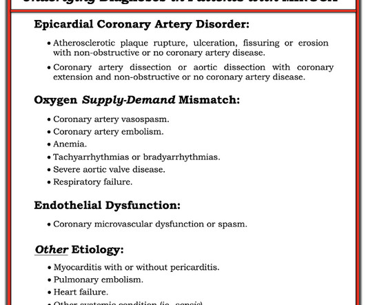
Category 1 : Sudden narrowing of a coronary artery due to ACS (plaque rupture with thrombosis and/or downstream showering of platelet-fibrin aggregates. It’s judicious, then, to arrange for coronary angiogram. Supply-demand mismatch (non-occlusive coronary disease, or exacerbation of preexisting flow insufficiency) a.
By Magnus Nossen This ECG is from a young man with no risk factors for CAD, he presented with chest pain. ACS then becomes less likely. Before the lab values returned this patient had a n emergent coronary CT angiogram done that ruled out CAD. Each main coronary artery (LAD, RCA and LCx) are shown in separate images.
The ECG is just a test: a Bayesian approach to acute coronary occlusion If a patient with a recent femur fracture has sudden onset of pleuritic chest pain, shortness of breath, and hemoptysis, the D-dimer doesn’t matter: the patient’s pre-test likelihood for PE is so high that they need a CT. A Bayesian approach to acute coronary occlusion.
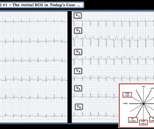
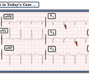
The biphasic T wave is consistent with recent reperfusion of an occluded coronary artery supplying the inferior region. Here’s the angiogram of the RCA : No thrombus or plaque rupture in the RCA (or any coronary artery) was found. This MI wasn’t caused by a ruptured plaque of CAD - it was a coronary artery dissection of the RCA.
A man in his 70s with past medical history of hypertension, dyslipidemia, CAD s/p left circumflex stent 2 years prior presented to the ED with worsening intermittent exertional chest pain relieved by rest. In our opinion it should not be given in ACS unless you are committed to the cath lab. 2009;95:1701–1706. Morris N, Body R.
A CT Coronary angiogram was ordered. Here are the results: --Minimally obstructive coronary artery disease. --LAD CAD-RADS category 1. --No Although a lesion is not visible anatomically on this CT scan, coronary catheter angiography could be considered based on Cardiology evaluation." A repeat troponin returned at 0.45
He had a history of CAD with CABG. Does this patient have ACS? He did not have ACS. Ventricular fibrillation is not only caused by acute coronary syndrome. The remainder were due to other etiologies, (including NonSTEMI ACS). But approximately 50% were due to non-ACS etiologies. The patient was cardioverted.
Sent by Anonymous, written by Pendell Meyers A man in his 60s with history of CAD and 2 prior stents presented to the ED complaining of acute heavy substernal chest pain that began while eating breakfast about an hour ago, and had been persistent since then, despite EMS administering aspirin and nitroglycerin. Pre-intervention.
The fire department, who operate at an EMT level in this municipality, arrived before us and administered 324 mg of baby aspirin to the patient due to concern for ACS. Just because you don't see hemodynamically significant CAD on angiogram does not mean it is not OMI. An angiogram is a "lumenogram;" most plaque is EXTRALUMINAL!!
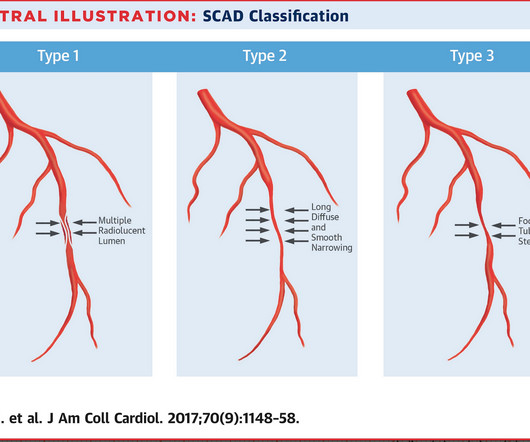
She had zero CAD risk factors. Now you have ECG and troponin evidence of ischemia, AND ventricular dysrhythmia, which means this is NOT a stable ACS. What is Spontaneous Coronary Artery Dissection (SCAD)? hours of substernal chest pressure. It was non-radiating and without other associated symptoms except for nausea.
Well, most commonly we’re going to see ACS. As the pregnant population continues to age and with RF and smoking and DM still common we can expect to see pregnant woman with CAD. Some unique features to consider in pregnancy is spontaneous coronary artery dissections.
But it does prove that the patient has coronary disease and makes the probability that his chest pain is due to ACS very very high. It is proven better than angiography alone in stable angina , and also has been shown to improve decisions on stenting non-culprit lesions in ACS. It could be acute, though probably is not.
Submitted and written by Alex Bracey with edits by Pendell Meyers and Steve Smith Case A 50ish year old man with a history of CAD w/ prior LAD MI s/p LAD stenting presented to the ED with chest pain similar to his prior MI, but worse. link] He was admitted to the cardiology unit for serial troponin measurements and concern for possible ACS.
A middle aged male with no h/o CAD presented with one week of crescendo exertional angina, and had chest pain at the time of the first ECG: Here is the patient's previous ECG: Here is the patient's presenting ED ECG: There is isolated ST depression in precordial leads, deeper in V2 - V4 than in V5 or V6. There is no ST elevation.
The diagnostic coronary angiogram identified only minimal coronary artery disease, but there was a severely calcified, ‘immobile’ aortic valve. Author continued : STE in aVR is often due to left main coronary artery obstruction (OR 4.72), and is associated with in-hospital cardiovascular mortality (OR 5.58).
Written by Pendell Meyers A man in his late 40s with several ACS risk factors presented with a chief complaint of chest pain. The cardiologists felt that the ECG did not represent ACS, and thought it was more likely pericarditis, so they did not take him to the cath lab. His first troponin T then resulted elevated at 0.19
The patient was transferred immediately for angiogram which revealed no significant CAD, and no intervention was performed. Coronary spasm causing massive current of injury with shark fin ECG. I would not expect ST-E to vanish in four beats with dissolving thrombus (also we know that the coronaries were clean).
He also had non-acute CAD of the RCA (50%) and LCX (50%). This is a h igher - p revalence H istory for acute coronary disease. Meyers : This ECG was texted to me with no clinical information, and my response was: "That looks like a very subtle LAD OMI. Cath images: Before intervention.
Furthermore, there was no family history of early CAD, MI, or sudden cardiac death. Cardiology admitted him for observation with plans for next-day coronary angiogram. He reported to EMS a medical history of GERD only. The physical exam was unremarkable for diaphoresis or pallor, and he denied any episodes of vomiting. 1] Driver, B.
Post by Smith and Meyers Sam Ghali ( [link] ) just asked me (Smith): "Steve, do left main coronary artery *occlusions* (actual ones with transmural ischemia) have ST Depression or ST Elevation in aVR?" Furthermore, among 35 patients with acute left main coronary artery occlusion, 9 presented with RBBB (mostly with LAFB) on the admission ECG.
If this is ACS with Aslanger's pattern , the ST depression vector of subendocardial ischemia (due to simultaneous 3 vessel or left main ACS) is directed toward lead II (inferior and lateral). Thus, this apparently is Aslanger's Pattern (inferior OMI with single lead STE in lead III, with simultaneous subendocardial ischemia).
The axiom of "type 1 (ACS, plaque rupture) STEMIs are not tachycardic unless they are in cardiogenic shock" is not applicable outside of sinus rhythm. This case represents the same physiologic event as OMI in terms of the result on the myocardium, therefore with identical ECG features, however there may not be ACS!
A middle-aged male with h/o CAD and stents presented with typical chest pressure. Is there likely to be fixed coronary stenosis that led to demand ischemia during pneumonia? --Was It is highly associated with proximal LAD occlusion or severe left main ACS and with bad outcomes. This is a very common misread.
She did not receive any opioids (which would mask her pain without affecting any underlying ACS). She also had non-acute CAD of the left main (50%) and LCX (75%). By the time the patient arrived at our facility, she had received aspirin and nitroglycerin, and her pain had apparently completely resolved. They opened it. Blondheim et al.
This page summarises the most current recommendations for the management of acute coronary syndromes with persistent ST-segment elevations (i.e III A Primary percutaneous coronary intervention strategy Management Recommendation Level of evidence Primary PCI of the infarct related artery (IRA) is indicated.
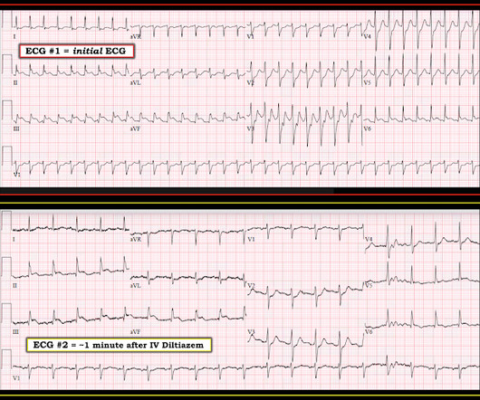
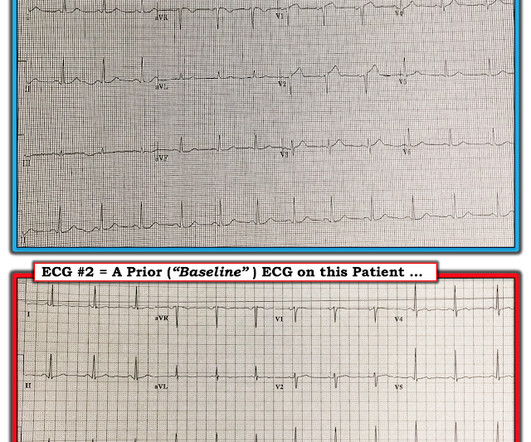
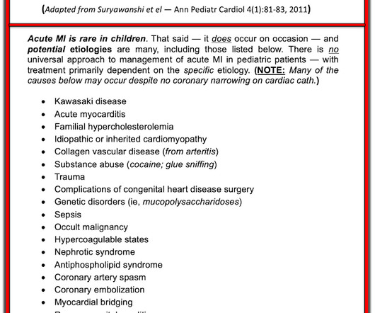
Acute coronary syndrome in a pediatric patient? He did have a family history notable for early CAD. A final ECG was perfomed on hospital day 2: Persistent ST elevation in the inferior leads with slight reciprocal ST depression in aVL Teaching points - It is essential to consider ACS in all age groups. Epub 2021 May 20.
A 70-something dialysis patient presented and coronary disease had missed dialysis and then presented with acute onset of shortness of breath. Negative trops and negative angiogram does not rule out coronary ischemia or ACS. These features are most likely the result of significant underlying coronary disease.
Case A 68 year old man with a medical history of hypertension, hyperlipidemia, and CAD with stent deployment in the RCA presented to the emergency department with chest pain. html ) Despite an undetectable troponin and three normal EKGs, the nature of the patients symptoms and his positive cardiac history warranted concern for ACS.
Written by Pendell Meyers A woman in her 70s with known prior coronary artery disease experienced acute chest pain and shortness of breath. But thankfully, when the clinical context is clearly and highly concerning for ongoing ischemia from ACS, this distinction doesn't matter much. Vital signs were within normal limits.
High-Sensitivity Cardiac Troponin I and Clinical Risk Scores in Patients With Suspected Acute Coronary Syndrome. Does a negative ECG and 2 undetectable troponins rule out ACS? CT Coronary angiogram The patient underwent CT Coronary Angiogram: Calcium score was 3600! Circulation [Internet] 2018;138(16):165465.
We organize all of the trending information in your field so you don't have to. Join 5,000+ users and stay up to date on the latest articles your peers are reading.
You know about us, now we want to get to know you!
Let's personalize your content
Let's get even more personalized
We recognize your account from another site in our network, please click 'Send Email' below to continue with verifying your account and setting a password.


Let's personalize your content