article thumbnail

Is this STEMI or NSTEMI? Neither. It is OMI.

Dr. Smith's ECG Blog

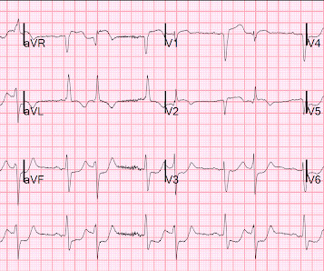
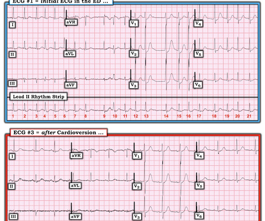
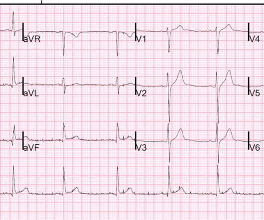
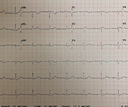
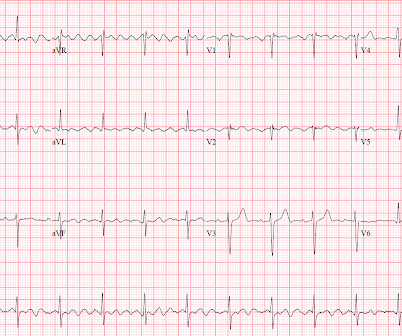
interesting spontaneous reperfusion case 1413140 prehospital STEMI first ED ECG is here, with 3/10 pain: But this is the same patient just 10 minutes before, with 7/10 pain Isn't it ridiculous to say that the patient has both a STEMI and an NSTEMI? ACS is dynamic. It can't be given one static name.

article thumbnail

What can you find with continuous ST monitoring in the ED?

Dr. Smith's ECG Blog

This dynamic change is diagnostic of ACS. Cardiology was consulted and agreed that his history was high risk for ACS and a next-day angiogram was merited. This was also non-diagnostic for OMI, although the dynamic changes are diagnostic of ACS. ECG at time 82 minutes: What do you think?

ED
professionals

Sign Up for our Newsletter

This site is protected by reCAPTCHA and the Google Privacy Policy and Terms of Service apply.

article thumbnail

What is the culprit artery? Not what you think.

Dr. Smith's ECG Blog

The history is highly suggestive of ACS. It is tempting to say that the ST depression was "posterior STEMI" on these initial ECGs, but that is not so. The ST depression was not V1-V3 (as in posterior STEMI), but rather V3-V6 (which is what is seen in subendocardial ischemia). STEMI and NonSTEMI exist on a spectrum.

article thumbnail

Chest Pain and Cardiogenic Shock with Profound ST Depression & STE in aVR. Activate the Cath Lab?

Dr. Smith's ECG Blog

It is important to note that these findings, if due to atherothrombotic acute coronary syndrome (ACS), are NOT due to occlusion of the left main, as is frequently stated in online postings and in literature. It is most commonly due to demand ischemia, not due to ACS! If it were ACS, what reperfusion options were available?

ACS
article thumbnail

repost this one as QRS distortion

Dr. Smith's ECG Blog

Some are STEMI-equivalents. Here are some basic concepts before we get into the ECGs: STEMI and NonSTEMI are arbitrary terms that may confuse the clinician. Inferior STEMI with AV Block, Cardiogenic Shock an. The QRS proves it. Posted by Steve Smith at 6:29 AM Email This BlogThis! Patient dies. Atrial Fib with RVR.

article thumbnail

A 60-something has chest tightness, palpitations, and ST depression V1-V3

Dr. Smith's ECG Blog

A prehospital ECG showed ST Depression in V1-V3 and the medics were concerned for posterior STEMI. Charts showed a history of some mitral regurgitation and an enlarge left atrium, as well as hypertension treated with a thiazide diuretic. Pulse was 128 and irregular, BP was 141/75.

ACS
article thumbnail

Interns are not (yet) poisoned by the STEMI/NSTEMI paradigm

Dr. Smith's ECG Blog

Now as an intern, he is exceptional at EKG interpretation because he was able to learn of the OMI paradigm and importance of pattern recognition before getting poisoned by years of learning STEMI. This is really a transient OMI (or transient STEMI if one uses that terminology and it has diagnostic ST Elevation).