This site uses cookies to improve your experience. To help us insure we adhere to various privacy regulations, please select your country/region of residence. If you do not select a country, we will assume you are from the United States. Select your Cookie Settings or view our Privacy Policy and Terms of Use.
Cookie Settings
Cookies and similar technologies are used on this website for proper function of the website, for tracking performance analytics and for marketing purposes. We and some of our third-party providers may use cookie data for various purposes. Please review the cookie settings below and choose your preference.
Used for the proper function of the website
Used for monitoring website traffic and interactions
Cookie Settings
Cookies and similar technologies are used on this website for proper function of the website, for tracking performance analytics and for marketing purposes. We and some of our third-party providers may use cookie data for various purposes. Please review the cookie settings below and choose your preference.
Strictly Necessary: Used for the proper function of the website
Performance/Analytics: Used for monitoring website traffic and interactions
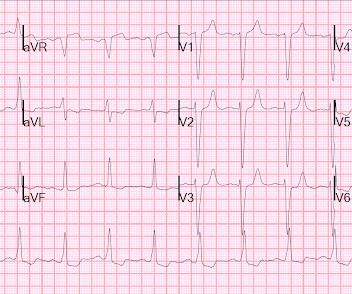
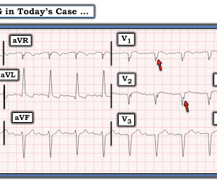
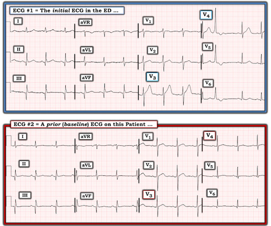
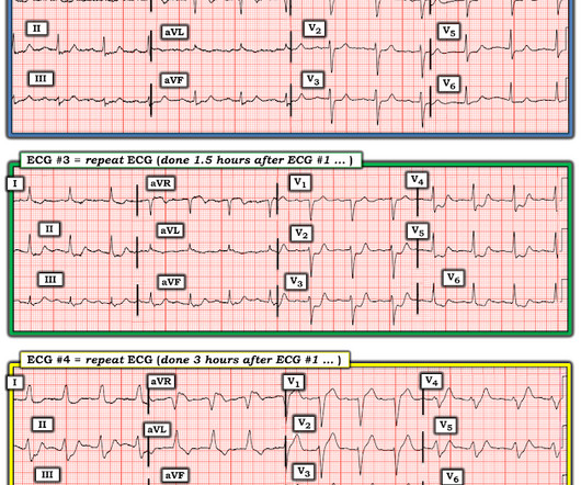
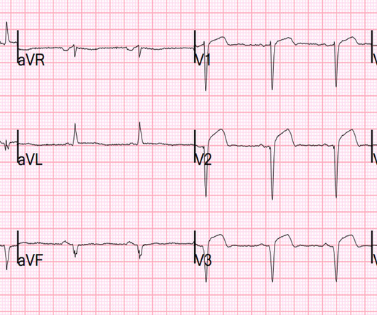
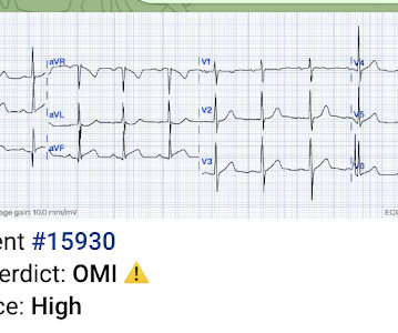
The T wave changes that have occurred are widespread, and not in a typical coronary distribution. Each time the patient underwent cardiac catheterization — and each time, she had patent coronary arteries! Discussion : ECG #2 shows sinus rhythm with quite dramatic change in T wave morphology when compared to ECG #1.
A 41-year-old South Asian male with history of hypertension, alcohol use disorder and hyperlipidemia, who has a strong family history of CAD presented with central substernal burning, pressure, and pain with associated diaphoresis. Coronary angiography before and after intervention is shown below. New PMcardio for Individuals App 3.0
An early and simple predictor of severe left main and/or three-vessel disease in patients with non-ST-segment elevation acute coronary syndrome. Because if such severe CAD is present, the patient is likely to need CABG. Am J Cardiol;107(4):495-500. why is this important?
A patient with history of severe CAD, CABG, with all native vessels occluded, on maximal medical therapy presented with his typical angina. NSTEMI: Patient with known severe CAD presenting with troponin elevation up to 21 and chest pain that was refractory to initial nitroglycerin therapy suggestive of unstable angina.
Denies family hx of coronary artery disease and premature cardiac death. Other DDX include, but less likely, coronary spasm, plaque rupture, and coronary microvascular dysfunction. Was taken to cath lab and no obstructive CAD was found. TTE with lateral WMA and reduced EF. LMCA: Normal.
Of course, any ECG (whether there is LBBB or normal conduction) can hide ischemia or even hide total acute coronary occlusion. Besides tachycardia, cardiomyopathy is also associated with excessive ST deviation. And patients with a previously poor ejection fraction cannot risk any infarction.
I think the right answer is that the patient probably needs emergent angiography to rule out acute coronary occlusion, but because it is such a complicated patient with such atypical symptoms, it is best to consult with cardiology about the case before activating. There was clearly a myocardial infarction and severe coronary disease.
There is increased LV cavity dimensions with an increase in transient ischemic dilation, suggesting Left Main, or 3-vessel coronary artery disease. 2. Coronary angiography reveals significant and severe CAD involving all three epicardial vessels. Strongly positive stress ECG Lexiscan administration. Type I ischemia.
Major adverse cardiac event rates in moderate-risk patients: Does prior coronary disease matter? Major adverse cardiac event rates in moderate-risk patients: Does prior coronary disease matter? He has no history of coronary artery disease. Date: June 30th, 2022 Reference: McGinnis et al. AEM June 2022. AEM June 2022.
He denied any known history of CAD, but did report ASCVD risk factors to include HTN, HLD, and DM. Ultimately the patient went to Cath and was found to have multi-vessel obstructive coronary disease with an acute LCX culprit vessel, which was stented. The patient was very uncomfortable, dyspneic, and displayed an SpO2 90% on RA.
A 63 year old man with a history of hypertension, hyperlipidemia, prediabetes, and a family history of CAD developed chest pain, shortness of breath, and diaphoresis after consuming a large meal at noon. They too have dense white masses consistent with coronary atherosclerosis. Edited by Smith He also sent me this great case.
By Magnus Nossen This ECG is from a young man with no risk factors for CAD, he presented with chest pain. Before the lab values returned this patient had a n emergent coronary CT angiogram done that ruled out CAD. Each main coronary artery (LAD, RCA and LCx) are shown in separate images. There are no coronary stenoses.
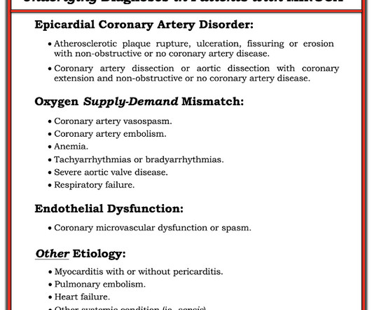
Category 1 : Sudden narrowing of a coronary artery due to ACS (plaque rupture with thrombosis and/or downstream showering of platelet-fibrin aggregates. It’s judicious, then, to arrange for coronary angiogram. Supply-demand mismatch (non-occlusive coronary disease, or exacerbation of preexisting flow insufficiency) a.
Moreover, he had no pertinent medical history to report in terms of CAD, HTN, HLD, or DM, for example. One cannot rely on this feature as a means of detecting changes – subtle, or dramatic – for volatile occlusive coronary thrombus. A 12 Lead ECG was recorded. Raw findings include Sinus Rhythm amidst an otherwise normal QRS.
The ECG is just a test: a Bayesian approach to acute coronary occlusion If a patient with a recent femur fracture has sudden onset of pleuritic chest pain, shortness of breath, and hemoptysis, the D-dimer doesn’t matter: the patient’s pre-test likelihood for PE is so high that they need a CT. A Bayesian approach to acute coronary occlusion.
A man in his mid 60s with history of CAD and stents experienced sudden onset epigastric abdominal pain radiating up into his chest at home, waking him from sleep. This patient in today's case was a man in his 60s with a known history of coronary disease, including prior stents. This is a re-post of an excellent case from 2021.
Angiogram: Severe two-vessel coronary artery disease with possible co-culprits (90% proximal circumflex, 70% mid/distal RCA) in the setting of non-ST elevation myocardial infarction. Marked ST depression from multi-vessel coronary disease serves to attentuate what would have been ST elevation in leads II and aVF ).
A CT Coronary angiogram was ordered. Here are the results: --Minimally obstructive coronary artery disease. --LAD CAD-RADS category 1. --No Although a lesion is not visible anatomically on this CT scan, coronary catheter angiography could be considered based on Cardiology evaluation." A repeat troponin returned at 0.45
He has a history of known CAD, diabetes, and dyslipidemia. The ED ECG in the context of the prehospital ECGs was indeed diagnostic of acute coronary occlusion. Cath Results: The cath lab was activated and co-culprit lesions were found: 99% circumflex and 95% right coronary artery (RCA). Both were stented.
The biphasic T wave is consistent with recent reperfusion of an occluded coronary artery supplying the inferior region. Here’s the angiogram of the RCA : No thrombus or plaque rupture in the RCA (or any coronary artery) was found. This MI wasn’t caused by a ruptured plaque of CAD - it was a coronary artery dissection of the RCA.
This is for the version housed on Telegram: [link] You can get the full PM Cardio app here if you live in the UK or EU (or say you do upon registration): [link] Case Continued The cath lab was activated and the patient received 180 mg of ticagrelor, and then was transported to the cath lab.
Angiography showed normal coronaries. MINOCA: Myocardial Infarction in the Absence of Obstructive Coronary Artery Disease). Here is my comment on MINOCA: "Non-obstructive coronary disease" does not necessarily imply "no plaque rupture with thrombus." 2) overlooked obstructive coronary disease (e.g., The K was normal.
No cardiac history but many risk factors for coronary disease. 5518881 A 60 yo F presented with new onset SOB over 1 week. She had new onset pulmonary edema with profound hypoxia. There was no previous ECG, echo, or angiogram available. Pulse was 108, RR 26, BP 120/91. Improved on BiPAP, furosemide.
He had a history of CAD with CABG. Ventricular fibrillation is not only caused by acute coronary syndrome. A middle-aged male had a V Fib arrest. He had not complained of any premonitory symptoms (which is very common). Here was his initial ED ECG: There is atrial fibrillation with a rapid ventricular response. He did not have ACS.
A man in his 70s with past medical history of hypertension, dyslipidemia, CAD s/p left circumflex stent 2 years prior presented to the ED with worsening intermittent exertional chest pain relieved by rest. The De Winter ECG pattern: morphology and accuracy for diagnosing acute coronary occlusion: systematic review. 2009;95:1701–1706.
I was called by Dr. YYYY from the Cath Lab - we reviewed the coronary anatomy. The patient had evidence of significant three-vessel disease, but an occluded right coronary artery. He was on high dose of pressors prior to arrival to the cardiac catheterization lab. Hence, decision made to ECMO cannulation prior to transfer.
This patient had known coronary artery disease (CAD), and previously required drug eluting stents to the obtuse marginal and diagonal arteries. 1 Despite the rarity of dextrocardia, coronary artery disease can occur with a similar frequency to that of the general population. Coronary heart disease in situs inversus totalis.
Hospital Course The patient was taken emergently to the cath lab which did not reveal any significant coronary artery disease, but she was noted to have reduced EF consistent with Takotsubo cardiomyopathy. Just because you don't see hemodynamically significant CAD on angiogram does not mean it is not OMI. It can only be seen by IVUS.
Sent by Anonymous, written by Pendell Meyers A man in his 60s with history of CAD and 2 prior stents presented to the ED complaining of acute heavy substernal chest pain that began while eating breakfast about an hour ago, and had been persistent since then, despite EMS administering aspirin and nitroglycerin. Pre-intervention.
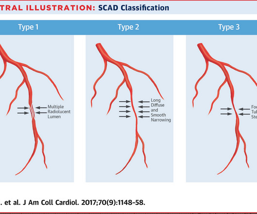
She had zero CAD risk factors. Next day, t he patient was taken for an angiogram and found to have a reperfused LAD lesion with good flow that appeared to the angiographer as if it was a spontaneous coronary artery dissection. What is Spontaneous Coronary Artery Dissection (SCAD)? hours of substernal chest pressure.
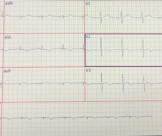
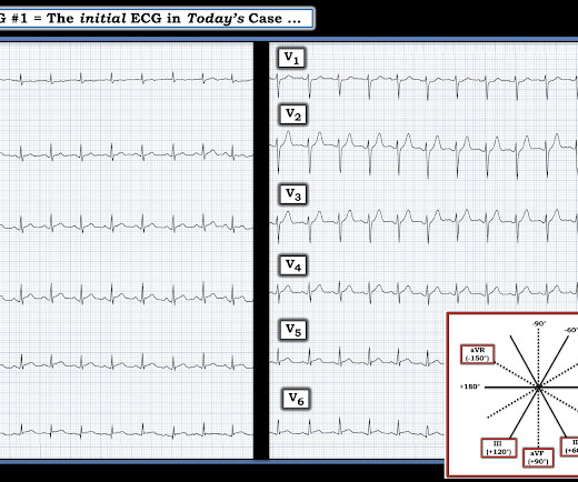
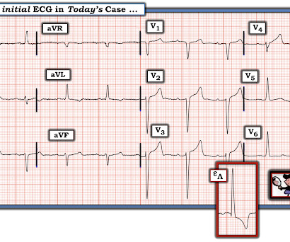
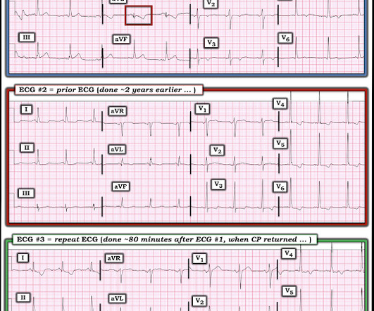
Concerning history, known CAD" Recorded 2 hours after pain onset: What do you think? To realize — Assessment of ECG #1 is complicated by knowing: i ) That today’s patient has a history of documented CAD ; and , ii ) The lack o f a prior tracing for comparison at the time the initial ECG was interpreted. What Do We Learn from ECG #3 ?
I want all to know that, with the right mind preparation, and the use of the early repol/LAD occlusion formula, extremely subtle coronary occlusion can be detected prospectively, with no other information than the ECG. It is not a missed STEMI, but it is a missed coronary occlusion. Wang T, Zhang M, Fu Y, et al.
As the pregnant population continues to age and with RF and smoking and DM still common we can expect to see pregnant woman with CAD. Some unique features to consider in pregnancy is spontaneous coronary artery dissections.
GLP-1 agonists are also associated with improved ejection fraction, coronary blood flow, and cardiac output while reducing the risk of cardiovascular events, infarction size, and all-cause mortality. Increased risk in those with preexisting CKD, other risk factors for renal disease (HTN or CAD), and those on ACEIs/ARBs.
But it does prove that the patient has coronary disease and makes the probability that his chest pain is due to ACS very very high. Instantaneous wave-free ratio is performed using high fidelity pressure wires that are passed distal to the coronary stenosis. Acute T-waves are large, even if not necessarily hyperacute.
The ED provider ordered a coronary CT scan to assess the patient for CAD. The patient was taken emergently to the cath lab for a pericardiocentesis instead of a coronary angiogram. Three months prior to this presentation, he received a pacemaker for severe bradycardia and syncope due to sinus node dysfunction.
A middle aged male with no h/o CAD presented with one week of crescendo exertional angina, and had chest pain at the time of the first ECG: Here is the patient's previous ECG: Here is the patient's presenting ED ECG: There is isolated ST depression in precordial leads, deeper in V2 - V4 than in V5 or V6. There is no ST elevation.
He had a family history of early CAD and occasional drug and tobacco use. However, subtle coronary occlusion may be completely missed by the computer and called "normal." It is not yet available, but this is your way to get on the list. link] Here is the history: A 30 yo man presented complaining of severe chest pain. References : 1.
The diagnostic coronary angiogram identified only minimal coronary artery disease, but there was a severely calcified, ‘immobile’ aortic valve. Author continued : STE in aVR is often due to left main coronary artery obstruction (OR 4.72), and is associated with in-hospital cardiovascular mortality (OR 5.58).
Is this due to coronary occlusion? The medic activated the cath lab but was refused by the interventionalist, who did not believe that this ECG represented acute coronary occlusion. But what we truly care about is coronary occlusion, for which STEMI is just a surrogate that is only about 75% sensitive for occlusion.
The patient was transferred immediately for angiogram which revealed no significant CAD, and no intervention was performed. Coronary spasm causing massive current of injury with shark fin ECG. I would not expect ST-E to vanish in four beats with dissolving thrombus (also we know that the coronaries were clean).
Case history A middle-aged woman with a history of HTN, but no prior CAD, presented to the ED with chest pain. LVH can mimic an acute anterior coronary occlusion (ACO) on the ECG. Electrocardiographic left ventricular hypertrophy in chest pain patients: Differentiation from acute coronary ischemic events. J Electrocardiol.
Submitted and written by Alex Bracey with edits by Pendell Meyers and Steve Smith Case A 50ish year old man with a history of CAD w/ prior LAD MI s/p LAD stenting presented to the ED with chest pain similar to his prior MI, but worse. Despite having acute coronary occlusion by cath, his ECGs never met STEMI criteria.
They found non-obstructive CAD, with only a 20% stenosis of OM2 and 10% RCA. A repeat ECG was performed and cardiology was re-consulted: Roughly unchanged. With the troponin elevated and ongoing pain, cardiology now decided to take him to the lab. No acute culprit. He was admitted to cardiology.
We organize all of the trending information in your field so you don't have to. Join 5,000+ users and stay up to date on the latest articles your peers are reading.
You know about us, now we want to get to know you!
Let's personalize your content
Let's get even more personalized
We recognize your account from another site in our network, please click 'Send Email' below to continue with verifying your account and setting a password.


Let's personalize your content