Remove CAD Remove Coronary Remove OR
article thumbnail

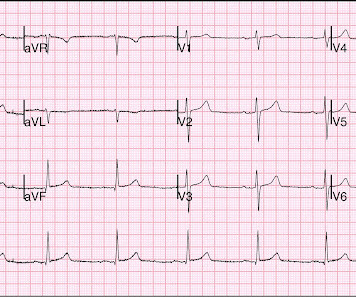
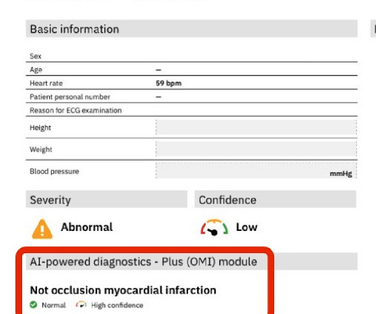
A 70 something female with chest pain (KG- done)

Dr. Smith's ECG Blog

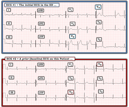
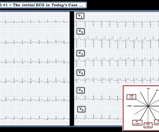
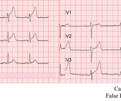
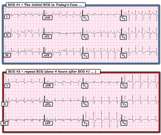
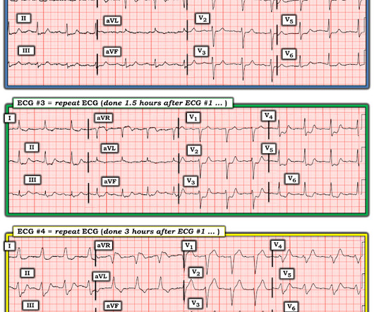
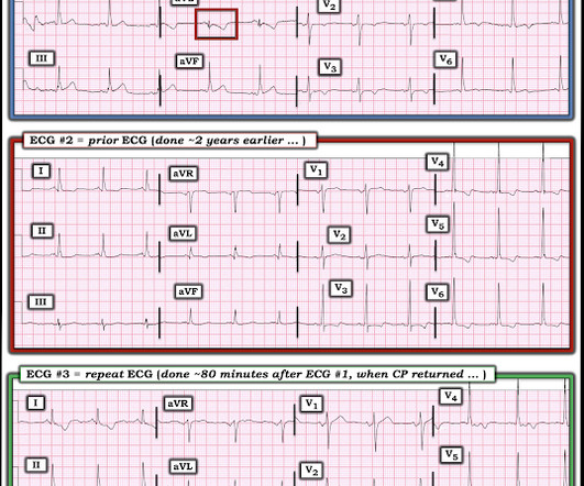
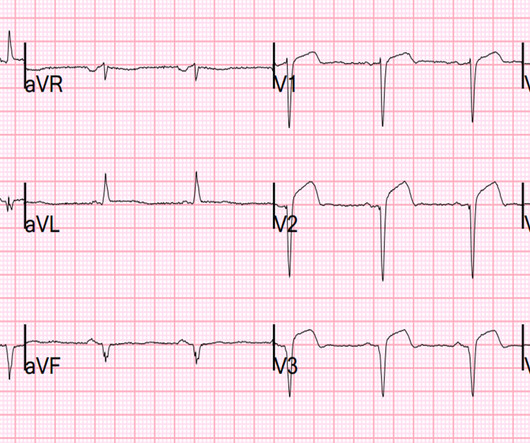
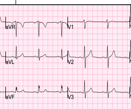
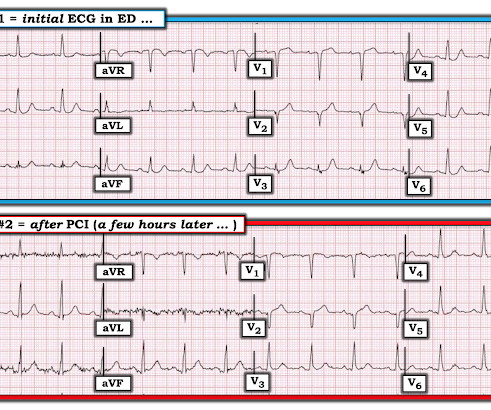
The T wave changes that have occurred are widespread, and not in a typical coronary distribution. Each time the patient underwent cardiac catheterization — and each time, she had patent coronary arteries! We proved this in this article. Note also the loss of R wave amplitude in ECG #2 compared to ECG #1. This patient never had ACS.

ACS 52
article thumbnail

Interns are not (yet) poisoned by the STEMI/NSTEMI paradigm

Dr. Smith's ECG Blog

A 41-year-old South Asian male with history of hypertension, alcohol use disorder and hyperlipidemia, who has a strong family history of CAD presented with central substernal burning, pressure, and pain with associated diaphoresis. Coronary angiography before and after intervention is shown below. Smith : a reperfused OMI is high risk.

STEMI 70
professionals

Sign Up for our Newsletter

This site is protected by reCAPTCHA and the Google Privacy Policy and Terms of Service apply.

article thumbnail

ST Elevation in aVR

Dr. Smith's ECG Blog

An early and simple predictor of severe left main and/or three-vessel disease in patients with non-ST-segment elevation acute coronary syndrome. Because if such severe CAD is present, the patient is likely to need CABG. Am J Cardiol;107(4):495-500. why is this important?

CAD 52
article thumbnail

STE aVR treated by me with high dose NTG, resolved, cath lab in AM, in ST depression ischemia folder

Dr. Smith's ECG Blog

A patient with history of severe CAD, CABG, with all native vessels occluded, on maximal medical therapy presented with his typical angina. NSTEMI: Patient with known severe CAD presenting with troponin elevation up to 21 and chest pain that was refractory to initial nitroglycerin therapy suggestive of unstable angina.

CAD 52
article thumbnail

LBBB: is there proportionally excessive discordant ST Elevation?

Dr. Smith's ECG Blog

Of course, any ECG (whether there is LBBB or normal conduction) can hide ischemia or even hide total acute coronary occlusion. What is difficult to remember is this: we excluded patients with a heart rate above 130 as tachycardia distorts the ST segments in LBBB. ECG 1 has a heart rate that is not above 130, but it is close. [We

CAD 52
article thumbnail

MINOCA case -- we need one of these and to discuss the topic

Dr. Smith's ECG Blog

Denies family hx of coronary artery disease and premature cardiac death. Other DDX include, but less likely, coronary spasm, plaque rupture, and coronary microvascular dysfunction. Was taken to cath lab and no obstructive CAD was found. TTE with lateral WMA and reduced EF. LMCA: Normal.

CAD 52
article thumbnail

Two Cases: Was it an error to activate the cath lab? Add AG case for 3rd one, except it is already listed as inferior aneurysm case.

Dr. Smith's ECG Blog

I think the right answer is that the patient probably needs emergent angiography to rule out acute coronary occlusion, but because it is such a complicated patient with such atypical symptoms, it is best to consult with cardiology about the case before activating. There was clearly a myocardial infarction and severe coronary disease.