This site uses cookies to improve your experience. To help us insure we adhere to various privacy regulations, please select your country/region of residence. If you do not select a country, we will assume you are from the United States. Select your Cookie Settings or view our Privacy Policy and Terms of Use.
Cookie Settings
Cookies and similar technologies are used on this website for proper function of the website, for tracking performance analytics and for marketing purposes. We and some of our third-party providers may use cookie data for various purposes. Please review the cookie settings below and choose your preference.
Used for the proper function of the website
Used for monitoring website traffic and interactions
Cookie Settings
Cookies and similar technologies are used on this website for proper function of the website, for tracking performance analytics and for marketing purposes. We and some of our third-party providers may use cookie data for various purposes. Please review the cookie settings below and choose your preference.
Strictly Necessary: Used for the proper function of the website
Performance/Analytics: Used for monitoring website traffic and interactions
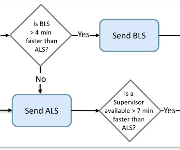
Some computer-aided dispatch (CAD) software did better than others by considering the average travel time of an actual route instead of allowing nearness to be determined by a straight-line distance. It was a matter of determining which crew was available closest to the scene. Life-threatening requests are similarly streamlined.
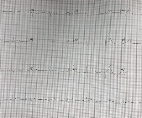
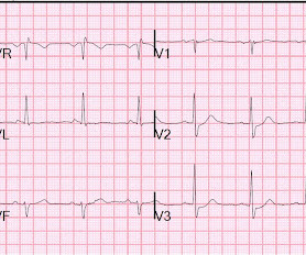
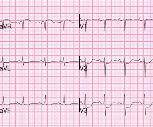
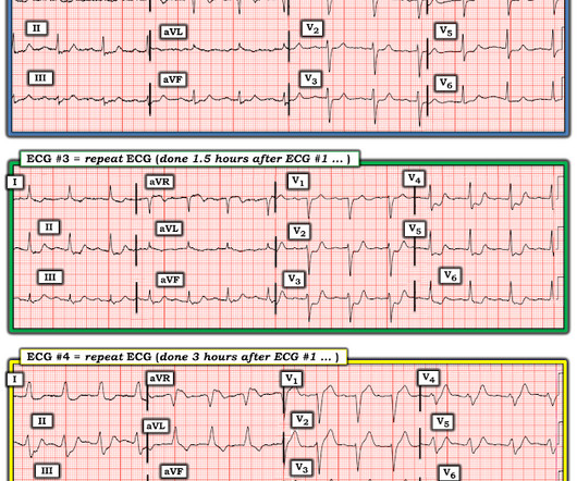
Patient 2 A man in his 50s with history of CAD and prior PCI, diabetes, presented with acute constant chest pain for the past few hours. Triage ECG: It was interpreted as lateral STEMI, and he was sent to the cath lab, where the angiogram showed unchanged CAD from known prior, with no acute culprit. He was discharged home.
2. Coronary angiography reveals significant and severe CAD involving all three epicardial vessels. He awoke earlier that morning in his usual state of health. His confusion progressively dissipated enroute to the hospital. At the time of ED arrival he was alert, oriented, and verbalizing only a headache with a normalized BP.
Spoon Feed — After a cervical artery dissection (CAD), patients with occlusive dissection had a reduced rate of subsequent ischemic stroke if treated with anticoagulation rather than antiplatelet treatment. We always work hard, but we may not have time to read through a bunch of journals. It’s time to learn smarter. N Engl J Med.
He has a history of CHF, dilated cardiomyopathy, HTN, HLD and CAD. These are very commonly encountered in the emergency department, so being able to correctly identify the rhythm is extremely important. Lets dive in! When you are presented with a tachycardic ECG, we want you to focus on two major factors right away. Take a look: Figure 3.
Connections with CAD, Billing, and Patient Outcomes Seeing the full picture of a patient’s care is key, and new technology is making the response for prehospital care faster and more efficient than ever. Real-time Access to Patient Data When every second counts, having the right information at the right time matters.
A man in his mid 60s with history of CAD and stents experienced sudden onset epigastric abdominal pain radiating up into his chest at home, waking him from sleep. This is a re-post of an excellent case from 2021. See it again now, along with our new Queen of Hearts functionality. We've come a long way in 2 years! And the pace only quickens.
Were making the jump to general availability (GA) and adding new features such as CAD and Cardiac Monitor integrations, Longitudinal Record (LR), and Mobile-to-Mobile functionality. This basic version will not include auto-import configuration, and integrations with CAD and EHR will not be added until upcoming releases in 2025.
A 63 year old man with a history of hypertension, hyperlipidemia, prediabetes, and a family history of CAD developed chest pain, shortness of breath, and diaphoresis after consuming a large meal at noon. Of course, writing “hypertensive emergency, underlying CAD with demand ischemia, or NSTEMI all remain on the differential” makes no sense.
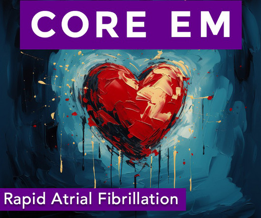
Metoprolol Considerations: Dosing (5 mg every 10-15 minutes, max 15 mg), benefits in CAD and HF, limitations in asthma/COPD patients. ECG Interpretation: Irregularly Irregular Rhythm: Absence of discernible P waves. Ventricular Rate: Typically over 100 bpm. Alternatives like procainamide or amiodarone are often more appropriate.
Because if such severe CAD is present, the patient is likely to need CABG. An early and simple predictor of severe left main and/or three-vessel disease in patients with non-ST-segment elevation acute coronary syndrome. Am J Cardiol;107(4):495-500. why is this important?
The patient presented to an outside hospital An 80yo female per triage “patient presents with chest pain, also hurts to breathe” PMH: CAD, s/p stent placement, CHF, atrial fibrillation, pacemaker (placed 1 month earlier), LBBB. HPI: Abrupt onset of substernal chest pain associated with nausea/vomiting 30 min PTA. This was stented with a 2.25
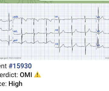
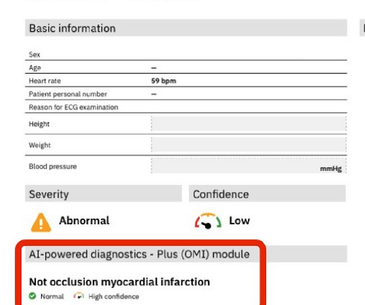
(THE PM CARDIO OMI AI APP) If you want this bot to help you make the early diagnosis of OMI and save your patient and his/her myocardium, you can sign up to get an early beta version of the bot here.
By Magnus Nossen This ECG is from a young man with no risk factors for CAD, he presented with chest pain. Before the lab values returned this patient had a n emergent coronary CT angiogram done that ruled out CAD. How would you assess this ECG? How confident are you in your assessment? What is your next step? How did the Queen do?
Once the new system is in place, it will also be able to integrate information from the computer-aided dispatch system (CADS). By Tom Jenkins Reprinted from the 2024 issue of Firefighter Strong In the fire academy, I doubt any candidate firefighter is excited or intrigued about incident reporting.
A patient with history of severe CAD, CABG, with all native vessels occluded, on maximal medical therapy presented with his typical angina. NSTEMI: Patient with known severe CAD presenting with troponin elevation up to 21 and chest pain that was refractory to initial nitroglycerin therapy suggestive of unstable angina.
In prior decades nearly all patients presenting to EDs with chest pain were admitted to hospital. If we thought about ACS, we brought them in. This would be for objective cardiac testing including stress test, CT-angiography, and/or invasive angiography. Major adverse cardiac event rates in moderate-risk patients: Does prior coronary disease matter?
Moreover, he had no pertinent medical history to report in terms of CAD, HTN, HLD, or DM, for example. CASE 1 A 45 y/o Male called 911 for new onset central chest discomfort, non-radiating, 5/10 pain scale, and without any vomiting, diaphoresis, or pallor. A 12 Lead ECG was recorded. There is no dramatic change, or evolution.
These concerns were readily conveyed to my supervising cardiologist with particular emphasis on high pretest probability for baseline advanced CAD (3-vessel disease, specifically) with a critically stenosed proximal LAD. This results in Type I MI. This results in Type II MI. What’s interesting is that the ECG can only detect ischemia.
She had a normal EF, and no significant CAD, and was taking flecainide to suppress the AF. Wide-complex tachycardia: VT or aberrant, or "other?" This case is contributed by Brooks Walsh , an EM physician and ECG expert from Connecticut. The case An older woman presented to the ED with dyspnea, diaphoresis, and chest pressure.
With API , participating CAD and RMS vendors will be able to automatically send data back and forth to NERIS. For any CAD and RMS vendors who are interested, you can share information and ask questions during the USFA development tea m’s regular NERIS office hours.
We hope to have a basic NERIS-compliant beta early in 2025, with additional updates (CAD imports, ESO EHR integration, ESO Insights reporting, etc.) It will be a simplified version and may not yet be ideal for agencies with ESO suite integrations like CAD and EHR imports. The timeline may vary. What will ESO charge for NERIS?
Similarly, if a patient with known CAD presents with refractory ischemic chest pain, the ECG barely matters: the pre-test likelihood of acute coronary occlusion is so high that they need an emergent angiogram. 1] European guidelines add "regardless of biomarkers".
Patient stated that he has had glucose over 400 even though he has not missed any doses of insulin. Aslanger's is a combination of inferior OMI with widespread ST depression and is due to BOTH occlusion of one artery (usually the circumflex, but sometimes the RCA) AND simultantous 3 vessel disease.
Angiogram The patient did go for an angiogram and there was no occlusion present, and no definite culprit, although there was diffuse CAD and one possible culprit: "There is a medium-large caliber OM3 with 40-50% stenosis in the mid-segment and lumen irregularity that could suggest dissection or plaque rupture, but TIMI III flow throughout."
Your existing historical CAD records contain the necessary information to build such dynamic views in real-time. The next step is to adequately distribute those available resources spatially to address the variation over the geographic area by time which requires an even deeper understanding of the call patterns.
This included date, POE, patient demographics, chief complaint, computer-aided dispatch (CAD), provider impression (diagnosis), and time out of service. Results : Over the study period, 8,407 encounters occurred at one of the three El Paso-Juárez POE, averaging 1,680 per year or 140 calls per month.
Transferred to our ED and taken to cath lab Cath- mild nonobstructive CAD, mild mid LAD bridge Echo- EF 65-70%, normal wall motion, no diastolic dysfunction. The cardiologist was skeptical of ACS according to his note. Sent to me: I looked at the EKG before reading any of your email. I thought it did not look like an OMI.
However, a smooth tapering of the mid-RCA was seen, highlighted in red below: How do we explain the MI if no sign of CAD was found? This MI wasn’t caused by a ruptured plaque of CAD - it was a coronary artery dissection of the RCA. Angiography Angiography was performed after aspirin and heparin were started.
Increased risk in those with preexisting CKD, other risk factors for renal disease (HTN or CAD), and those on ACEIs/ARBs. The effects of GLP-1 agonists are associated with the dose. Higher doses of GLP-1 agonists are associated with weight loss. Take for example semaglutide. Ozempic is utilized for DM2 in doses of 0.5, mg SQ every week.
He had no previous history of CAD, and presented with very typical waxing and waning chest pain, much worse with exertion but also present at rest and on presentation, though his pain was minimal at the time of the ECG. I saw this 59 year old male 3 weeks ago. Blood pressure was 150/80. The ECG normalized overnight. Maximum troponin was 2.1
Sent by Anonymous, written by Pendell Meyers A man in his 60s with history of CAD and 2 prior stents presented to the ED complaining of acute heavy substernal chest pain that began while eating breakfast about an hour ago, and had been persistent since then, despite EMS administering aspirin and nitroglycerin. Pre-intervention.
In my experience — even when everything points to Stress Cardiomyopathy — it is not always possible to rule out concomitant severe CAD, or even ACS. Smith comment : Acute posterior infarction most often does NOT have increased R-waves or early RS transition and this common belief is an obstacle to the diagnosis of acute posterior OMI.
This was a middle aged female with a h/o CAD who presented to the ED by EMS sudden onset of central chest pressure 45 min prior to ED arrival with associated diaphoresis and SOB. There is LVH and there are ST-T abnormalities (large inferior T-waves and ST elevation, with reciprocal findings in aVL).
Diagnosis of MINOCA should be made according to the Fourth Universal Definition of MI, in the absence of obstructive coronary artery disease (CAD) (no lesion ≥50%). The authors recommend using optical coherence tomography or intravascular ultrasound imaging in patients with evidence of nonobstructive CAD by angiogram. The K was normal.
He had a history of CAD with CABG. A middle-aged male had a V Fib arrest. He had not complained of any premonitory symptoms (which is very common). Here was his initial ED ECG: There is atrial fibrillation with a rapid ventricular response. There is profound ST depression especially in I, II, V2-V6. One should wait a short time (15 minutes?)
Multivessel CAD 2. My blind reading of the ECG (no clinical info): Severe ischemia, most consistent with occlusion of branch of the circumflex, but could also be subendocardial ischemia Chest X-ray i mpression: Findings suggest mild interstitial pulmonary edema. Underlying right basilar atelectasis and or infiltrate cannot be excluded.
No known risk factors for ACS/CAD. A medic sent this case A 33 yom who presented to an urgent care facility with a complaint of chest pain for several days. His chest pain started after he began taking a testosterone supplement. This is a classic case of benign T-wave inversion. There may or may not be LVH here.
As the pregnant population continues to age and with RF and smoking and DM still common we can expect to see pregnant woman with CAD. Critical illness in pregnancy is remarkably rare given the somewhat bonkers system for reproduction that we seem to have evolved over the past million or years. Improved care of complex.
A middle aged male with no h/o CAD presented with one week of crescendo exertional angina, and had chest pain at the time of the first ECG: Here is the patient's previous ECG: Here is the patient's presenting ED ECG: There is isolated ST depression in precordial leads, deeper in V2 - V4 than in V5 or V6. There is no ST elevation.
He had a family history of early CAD and occasional drug and tobacco use. This was sent by : Jacob Smith, DO Emergency Medicine Resident Ohio Health Doctors Hospital Emergency Residency Christopher Lloyd, DO, FACEP Director of Clinical Education, USACS Midwest Case A 30 year old patient presents to triage with chest pain.
The ED provider ordered a coronary CT scan to assess the patient for CAD. Three months prior to this presentation, he received a pacemaker for severe bradycardia and syncope due to sinus node dysfunction. At around 1430, as the patient was being prepared to leave for the scan, he developed severe chest pain, dizziness, and became diaphoretic.
A man in his 70s with past medical history of hypertension, dyslipidemia, CAD s/p left circumflex stent 2 years prior presented to the ED with worsening intermittent exertional chest pain relieved by rest. Written by Nathanael Franks MD, reviewed by Meyers, Smith, Grauer, etc. He was diagnosed as NSTEMI.
We organize all of the trending information in your field so you don't have to. Join 5,000+ users and stay up to date on the latest articles your peers are reading.
You know about us, now we want to get to know you!
Let's personalize your content
Let's get even more personalized
We recognize your account from another site in our network, please click 'Send Email' below to continue with verifying your account and setting a password.


Let's personalize your content