This site uses cookies to improve your experience. To help us insure we adhere to various privacy regulations, please select your country/region of residence. If you do not select a country, we will assume you are from the United States. Select your Cookie Settings or view our Privacy Policy and Terms of Use.
Cookie Settings
Cookies and similar technologies are used on this website for proper function of the website, for tracking performance analytics and for marketing purposes. We and some of our third-party providers may use cookie data for various purposes. Please review the cookie settings below and choose your preference.
Used for the proper function of the website
Used for monitoring website traffic and interactions
Cookie Settings
Cookies and similar technologies are used on this website for proper function of the website, for tracking performance analytics and for marketing purposes. We and some of our third-party providers may use cookie data for various purposes. Please review the cookie settings below and choose your preference.
Strictly Necessary: Used for the proper function of the website
Performance/Analytics: Used for monitoring website traffic and interactions
Time-sensitive emergencies are demanding events. Whether it's a STEMI, stroke, trauma incident—or even a mass casualty incident like a hurricane or multi vehicle accident—treatment time is often a dominant factor when determining the post-treatment wellbeing of a patient.
The subsequent diagnosis of an ST-segment elevation myocardial infarction (STEMI) sets forth a cascade of events that typically culminates in the patient being transported to hospital capable of emergent percutaneous coronary intervention (PCI). However, the notion of “STEMI equivalents” has gained traction.
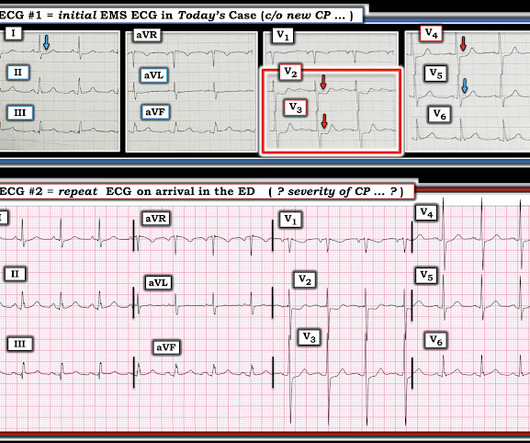
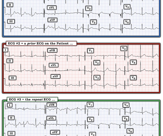
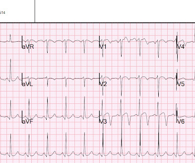
He had ongoing pain following the syncopal event but went to bed and awoke in the morning with ongoing pain. The first EKG was concerning for a Wellen’s-like pattern of subtle reperfusion changes in the setting of stuttering anginal-equivalent symptoms, but none were diagnostic of STEMI or OMI.
Major adverse cardiac events 40 minutes after giving the antiarrhythmic. The following table shows their results, take a look: Note that procainamide leads with less adverse cardiac events. Procainamide therapy was associated with less major cardiac adverse events and a higher proportion of tachycardia termination within 40 min.
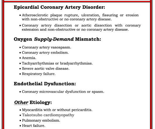
Patient assessments should include a complete history of both the event and patient’s medical problems. ST-segment Elevation Myocardial Infarction (STEMI) criteria have been the traditional method of cath lab activation. A recent meta-analysis showed that STEMI criteria are only 43% sensitive for occlusion myocardial infarction (OMI).
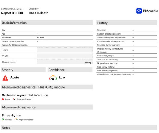
The Zoll algorithm impressively stated: STEMI A paramedic student had the PMCardio AI Queen of Hearts on his phone and this is what it reported: New PMcardio for Individuals App 3.0 Knowing this patient's age and his history of known coronary disease — immediately places him in a higher -risk group for an acute coronary event.
As the front line of the healthcare system, EMS delivers time-sensitive care in moments of crisis, bridging the gap between life-threatening events and definitive treatment. 1 EMS has always been described as the canary in the coal mine and the tip of the spear simultaneously. References 1. Emergency Medical Services Systems Development.
June Recap Celebrating Customer and Community Successes In Colorado Teams in Colorado Springs, CO, are using Pulsara to keep STEMI DTB times under 30 minutes, close feedback loops, and create a culture of transparency and trust across the entire emergency care continuum.
The time dependent emergencies, major trauma, STEMI, and stroke, do. Anecdotal reports may tug at the heartstrings, but our story would be so much more convincingly conveyed if we said that we had a 23% reduction in deaths from STEMI or stroke because of the system of care we have put into place.
He described the sensation and the sequence of events as a “Hampir Stroke” (Hampir stroke in the Malay Language can be literally translated “Near Stroke,” but colloquially probably means a near catastrophic event, like a Myocardial Infarction.) There were two other small scale events but all three were associated with fever.
The PMCardio Queen of Hearts app asks you, before giving an interpretation of OMI ("STEMI-Equivalent"), whether the patient's clinical presentation is high risk for OMI. Dizziness is so unlikely to be OMI without an obvious ECG, that I am going to pretend that this patient presented with chest pain. In the November 27, 2024 post Drs.
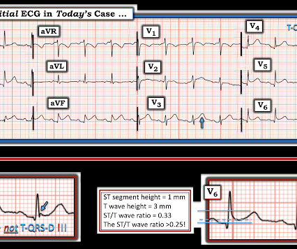
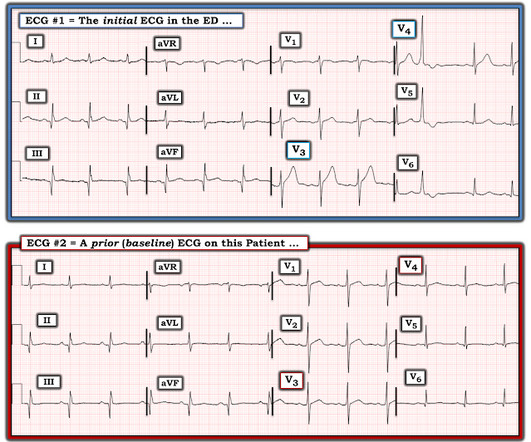
Without seeing the patient, my interpretation of the first ECG was: likely normal variant ST-elevation (early repolarization), with a small possibility of pericarditis, and almost no possibility of acute coronary occlusion (STEMI). and therefore highly unlikely to be STEMI. Mostly the same findings and reasoning as the current ECG.
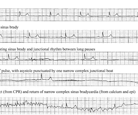
1] In a review of hyperkalemia in the ED, all adverse events occurred prior to calcium and usually with multiple signs of hyperkalemia (like the first ECG) with the biggest predictors not only wide QRS, but also bradycardia and/or junctional rhythm.[2] regardless of the ECG (when the repeat level came back).[1] References 1. Lindner et al.
Most of you will recall the JC list of February 2025, where a large Swedish study showed no association with neuropsychiatric adverse events of montelukast in children. Acute Intraoperative STEMI in a Toddler. This was most pronounced when concomitant antihistamines were used. J Child Neurol. 2025 Jun 2:8830738251328423. 2025 Jun 16.
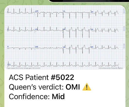
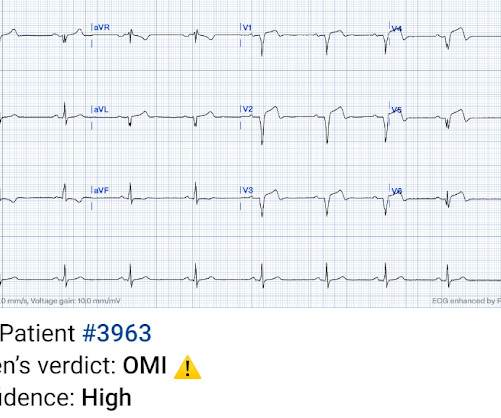
This ECG was recorded 30 minutes later with ongoing pain: Queen of Hearts says no signs of STEMI or Equivalent for both: New PMcardio for Individuals App 3.0 Midwest STEMI Consortium: decreased false positives by 33% (presented at ACC Quality Summit San Antonio 2024) 3. She denied preceding symptoms or recent illnesses.
Theres ST elevation in V3-4 which meets STEMI criteria, which could be present in either early repolarization, pericarditis or injury. Lets see what happens in the current STEMI paradigm. Emergency physician: STEMI neg but with elevated troponin = Non-STEMI The first ECG was signed off. What do you think?
She knows the baseline is normal, and she knows the STEMI(-) OMI one is diagnostic of OMI, with the highest possible confidence. Here is the EM decision making: "The patient's EKG revealed some repolarization abnormalities but no clear signs of a STEMI. Back to the case: Unfortunately, the ECG was not understood by the provider.
Obvious infero-postero-lateral STEMI(+)OMI, regardless of context Now let’s put them in order: what was the sequence? With serial ECGs that are ‘STEMI negative’ the physician could have waited for serial troponin levels or referred the patient as “non-STEMI”. What was the outcome and final diagnosis?
Methods and Results Patients with confirmed ST elevation myocardial infarction (STEMI) treated by emergency medical services were included in this retrospective cohort analysis of the AVOID study. There was no association between moderate to severe chest pain on arrival and major adverse cardiac events at 6 months (20% vs. 14%, p=0.12).
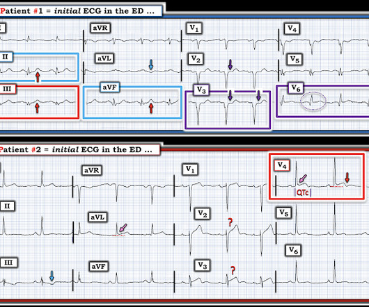
A prehospital “STEMI” activation was called on a 75 year old male ( Patient 1 ) with a history of hyperlipidemia and LAD and Cx OMI with stent placement. The two cases were considered: Patient 1 was recognized by the ED provider and the cardiologist as having resolved “STEMI”. He wrote most of it and I (Smith) edited.
So this NSTEMI was likely a STEMI(-)OMI with delayed reperfusion. The patient was admitted as ‘NSTEMI’ which is supposed to represent a non-occlusive MI, but the underlying pathophysiology is analogous to a transient STEMI. See these posts: Chest Pain, ST Elevation, and an Elevated Troponin: Should we Activate the Cath Lab?
Jason was very skeptical of STEMI. This also argues against STEMI. KEY POINTS from this CASE: The presenting history often provides invaluable clues to the likelihood of an acute cardiac event. ( This is a "low prevalence" history for an acute cardiac event.). He complained of 3 days of diarrhea and abdominal pain.
Their OMI Manifesto details how use of standard STEMI criteria results in an unacceptable level of inaccuracy, in which an estimated 25-30% of acute coronary occlusions are missed! The more leads with suspicious findings — the greater the concern for an acute ongoing event.
They wanted to know if I would like them to activate the outside hospital's "STEMI alert." But of course, this is not a STEMI by definition as it does not meet STEMI criteria. The STEMI guidelines do state that hyperacute T-waves "may indicate early acute myocardial infarction" but do not discuss it as a "STEMI equivalent."
Subtle as a STEMI." (i.e., Here is the bottom line of the article: It is widely believed that hyperacute T-waves are a transitional state preceding ST Elevation 1–4 Thus, it is tempting to postulate that early cases of OMI will eventually evolve to STEMI; yet, our data contradicts that notion. This one is easy for the Queen.
Even in patients whose moderate stenosis undergoes thrombosis, most angiograms show greater than 50% stenosis after the event. See "Prevention of cardiovascular disease events in those with established disease (secondary prevention) or at very high risk".) From Gue at al.
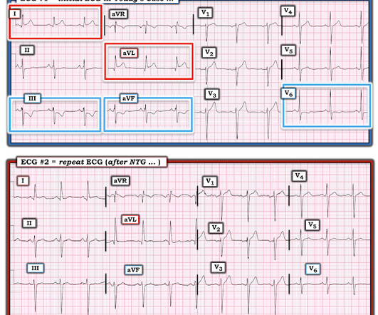
There’s inferior ST depression which is reciprocal to subtle lateral convex ST elevation, and the precordial T waves are subtly hyperacute – all concerning for STEMI(-)OMI of proximal LAD. There’s ST elevation I/aVL/V2 that meet STEMI criteria. This is obvious STEMI(+)OMI of proximal LAD. Non-STEMI or STEMI(-)OMI?
This patient could have very easily been overlooked, both because the ECG was STEMI negative and because the Q waves were attributed to an “old infarct”. Fortunately, Dr. Cho was not looking for STEMI ECG criteria but for an acute coronary occlusion. OMI or STEMI? As cardiology documented, “possible STEMI.
The cardiologist recognized that there were EKG changes, but did not take the patient for emergent catheterization because the EKG was “not meeting criteria for STEMI”. Troponin was elevated and no “STEMI” was seen on the EKG, so if it is acute MI, then “NSTEMI” is the diagnosis (however flawed), not a pathology on the differential.
This was a machine read STEMI positive OMI. The meaning of this quote is that at times, something as obvious as the dramatic anterior lead ST elevation that we see in today's tracing is not the result of an acute LAD STEMI. His ECG is shown below. Pretty obvious anterior current of injury. What would you guess is the culprit artery?
It does, in fact, the STE meets STEMI criteria since there is 1 mm of in V4 and V5. As discussed above in Dr. Smith's excellent discussion — serial ECGs, correlated to severity of patient symptoms soon confirmed the acute event in today's patient. This ECG was texted to me with no other information. What did I say?
It has been estimated that in the aggregate, they occur at a rate of about 3 per 1000 patients with acute MI, and most of these events occur in patients with STEMI. A mong patients with STEMI, ventricular septal rupture is the most common and free wall rupture is the least common.
Quiz : What percent of full blown STEMI have an open artery with normal flow at angiogram? It too is "normal" and you decide that this is not OMI or STEMI and you just decide to get troponins. So despite the artifact — and even without any history — this initial ECG has to be interpreted as an acute event until proven otherwise.
Troponin T peaked at 2074 ng/L (very high, typical of OMI/STEMI). As a result — the onset of any acute event that may have occurred is uncertain. Post PCI the patient became gravely hypotensive and "shocky". She stabilized on dobutamine and levosimendan infusions that could be discontinued after 24 hours. 21, 2017 ).
Here it is: Obvious Inferior Posterior STEMI (+) OMI. Initial troponin was: 3 ng/L We showed that the first troponin in acute STEMI is often negative in at least 27%. Aside on ECG Research: 20% of Definite diagnostic STEMI (Cox et al.) The cath lab was activated prehospital But imagine if the patient had walked in.
This is diagnostic of infero-posterior OMI, but it is falsely negative by STEMI criteria and with falsely negative posterior leads (though they do show mild ST elevation in V4R). They were less likely to have STEMI on ECG, and more likely to be initially diagnosed as non-ACS.
Smith : there is some minimal ST elevation in V2-V6, but does not meet STEMI criteria. Transient STEMI has been studied and many of these patients will re-occlude in the middle of the night. And dynamic ST-T wave changes between ECG #1 and ECG #2 confirm an acute event in progress. Is it normal STE? This is a "Transient OMI".
Only very slight STE which does not meet STEMI criteria at this time. I am immediately worried that this OMI will not be understood, for many reasons including lack of sufficient STE for STEMI criteria, as well as the common misunderstanding of "no reciprocal findings" which is very common with this particular pattern.
Patient still not having chest pain however this is more concerning for OMI/STEMI. Wellens' syndrome is a syndrome of Transient OMI (old terminology would be transient STEMI). As far as I can tell, there is only one randomized trial of immediate vs. delayed intervention for transient STEMI. Labs ordered but not yet drawn.
would require the ST/S ratio to be 25% for diagnosis of STEMI in LVH. The physician was concerned about STEMI, but also worried that she was overreacting, with the potential that LVH was producing a "STEMI-mimic." Can you diagnose an ACO (STEMI) when you also have LVH? The criteria of Armstrong et al. References 1.
Post Cath ECG: Obviously completing MI with LVA morphology, and STE that meets STEMI criteria (but pt is still diagnosed as "NSTEMI"). Day 12 ECG: FINAL DIAGNOSIS: "NSTEMI" Despite the fact that his day 4 ECG easily meets STEMI criteria, the patient is diagnosed as NSTEMI. No TIMI flow was listed in the report.
Here is his ED ECG at triage: Obvious high lateral OMI that does not quite meet STEMI criteria. Furthermore, if this occurs at all, it is a rare event. He does have a recently diagnosed PE, and has not been taking his anticoagulation due to cost. He had a previous ECG on file: Proving the findings are new The cath lab was activated.
It was tested on a large database of known outcomes and was more than twice as senstivity as STEMI criteria and much better than cardiologists. These are identical to Wellens' waves because Wellens' syndrome is a transient OMI (or transient STEMI) Learning Points 1. This is the first version of the AI system.
We organize all of the trending information in your field so you don't have to. Join 5,000+ users and stay up to date on the latest articles your peers are reading.
You know about us, now we want to get to know you!
Let's personalize your content
Let's get even more personalized
We recognize your account from another site in our network, please click 'Send Email' below to continue with verifying your account and setting a password.


Let's personalize your content