This site uses cookies to improve your experience. To help us insure we adhere to various privacy regulations, please select your country/region of residence. If you do not select a country, we will assume you are from the United States. Select your Cookie Settings or view our Privacy Policy and Terms of Use.
Cookie Settings
Cookies and similar technologies are used on this website for proper function of the website, for tracking performance analytics and for marketing purposes. We and some of our third-party providers may use cookie data for various purposes. Please review the cookie settings below and choose your preference.
Used for the proper function of the website
Used for monitoring website traffic and interactions
Cookie Settings
Cookies and similar technologies are used on this website for proper function of the website, for tracking performance analytics and for marketing purposes. We and some of our third-party providers may use cookie data for various purposes. Please review the cookie settings below and choose your preference.
Strictly Necessary: Used for the proper function of the website
Performance/Analytics: Used for monitoring website traffic and interactions
This was sent by an undergraduate (not yet in medical school, but applying now) who works as an ED technician (records all EKGs, helps with procedures, takes vital signs) and who reads this blog regularly. Diagnosis of Type I vs. Type II Myocardial Infarction in EmergencyDepartment patients with Ischemic Symptoms (abstract 102).
Written by Jesse McLaren A 70 year old with prior MIs and stents to LAD and RCA presented to the emergencydepartment with 2 weeks of increasing exertional chest pain radiating to the left arm, associated with nausea. 1] European guidelines add "regardless of biomarkers".
PEARL #2: = Aslanger's Pattern: Examples of Aslanger's Pattern appear in a number of cases in Dr. Smith's ECG Blog ( This pattern is very nicely described by Dr. Smith in the January 4, 2021 post). Patient stated that he has had glucose over 400 even though he has not missed any doses of insulin.
Upon arrival to the emergencydepartment, a senior emergency physician looked at the ECG and said "Nothing too exciting." Just because you don't see hemodynamically significant CAD on angiogram does not mean it is not OMI. I could have told you this (and did tell you this) without an MRI.
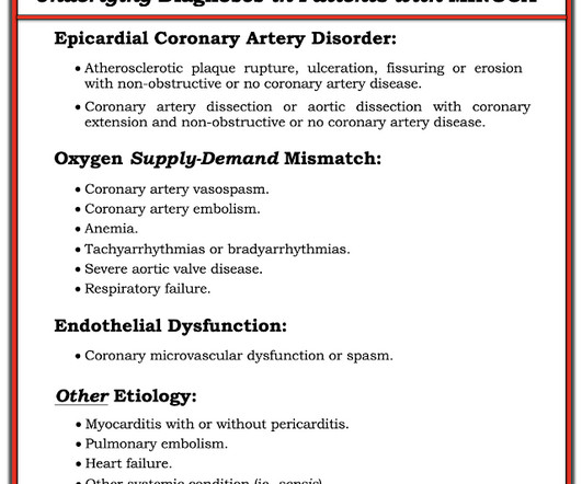
She was unable to be defibrillated but was cannulated and placed on ECMO in our EmergencyDepartment (ECLS - extracorporeal life support). Diagnosis of MINOCA should be made according to the Fourth Universal Definition of MI, in the absence of obstructive coronary artery disease (CAD) (no lesion ≥50%). myocarditis).
I finished my residency of Emergency Medicine and I’m working at a great EmergencyDepartment here in Brazil. So, I'm a follower of your blog, and I think I have a interesting case that I attended yesterday." Aguiar last week: "Last year I had a couple of lessons with you while on my internship.
Submitted and written by Alex Bracey with edits by Pendell Meyers and Steve Smith Case A 50ish year old man with a history of CAD w/ prior LAD MI s/p LAD stenting presented to the ED with chest pain similar to his prior MI, but worse. In Figure-1 I have excerpted from the above blog post, the Pearls of Wisdom from Drs.
So I went to look at the chart and here is the history: This patient with no h/o CAD had a couple of episodes of chest pain during the day, then presented with one hour of substernal chest pain that had some reproducibility but also improved from 10/10 to 5/10 with nitroglycerine. Ann Emerg Med 1998;31(1):3-11.
Written by Pendell Meyers A man in his late 30s with history of hypertension, tobacco use, and obesity presented to the EmergencyDepartment for acute chest pain which started approximately 3 hours prior to arrival, in the setting of a very stressful situation. Scattered other nonobstructive CAD.
At the bottom of the post, I have re-printed the section on aVR in my article on the ECG in ACS from the Canadian Journal of Cardiology: New Insights Into the Use of the 12-Lead Electrocardiogram for Diagnosing Acute Myocardial Infarction in the EmergencyDepartment Case 1. This was a 100% acute LM occlusion.
Written by Jesse McLaren, with comments from Smith An 85 year old with a history of CAD presented with 3 hours of chest pain that feels like heartburn but that radiates to the left arm. Below is the ECG. What do you think? There’s sinus bradycardia, first degree AV block, normal axis, delayed R wave progression, and normal voltages.
Case A 68 year old man with a medical history of hypertension, hyperlipidemia, and CAD with stent deployment in the RCA presented to the emergencydepartment with chest pain. Also : See Ken Grauer's excellent comments at the bottom. He developed it only 20 minutes prior to presentation while cutting branches outside.
She presented to the EmergencyDepartment at around 3.5 The procedure was described as very complex due to severe multivessel CAD, but ultimately PCI was successfully performed to the ostial LCX. This ECG is taken from the July 25, 2024 post in Dr. Smith's ECG Blog. Vital signs were within normal limits. Pre-intervention.
Undetectable high-sensitivity cardiac troponin T level in the emergencydepartment and risk of myocardial infarction. Many authors state that if troponins are undetectable and ECG is normal, no further testing should be done. For example, in JACC: Bandstein N, Ljung R, Johansson M, Holzmann MJ.
We organize all of the trending information in your field so you don't have to. Join 5,000+ users and stay up to date on the latest articles your peers are reading.
You know about us, now we want to get to know you!
Let's personalize your content
Let's get even more personalized
We recognize your account from another site in our network, please click 'Send Email' below to continue with verifying your account and setting a password.


Let's personalize your content